Electron microscopy of stretch-grown axons. Scanning electron
4.5 (358) · € 21.00 · En Stock

Download scientific diagram | Electron microscopy of stretch-grown axons. Scanning electron micrographs illustrating a small fascicle composed of axons 100-250 nm in diameter (A, B). Fasciculation of axons occurs during the elongation process as smaller bundles and individual axons coalesce and adhere to one another, forming larger bundles similar to the one depicted here. Transmission electron micrograph of cross sections near the center of axon fascicles in nonstretch conditions ( C) and axons stretched to a length of 5 cm in 14 d (D), showing no change in axon cytoskeletal structures. Scale bars: A, 10 m; B, 1 m; C, D, 500 nm. from publication: Extreme Stretch Growth of Integrated Axons | Large animals can undergo enormous growth during development, suggesting that axons in nerves and white matter tracts rapidly expand as well. Because integrated axons have no growth cones to extend from, it has been postulated that mechanical forces may stimulate axon | Axons, Growth Cones and Afferent Neurons | ResearchGate, the professional network for scientists.

Micromachines, Free Full-Text

Incidental Ultrastructural Findings in the Sural Nerve and Dorsal Root Ganglion of Aged Control Sprague Dawley Rats in a Nonclinical Carcinogenicity Study - William A. Meier, Michael J. Linn, Wendell P. Davis

Micromachines, Free Full-Text

Transmission Electron Microscopy and Morphometry of the CNS White Matter

Three dimensional electron microscopy reveals changing axonal and myelin morphology along normal and partially injured optic nerves

Wallerian Degeneration in the Optic Nerve Stretch-Injury Model of Traumatic Brain Injury: A Stereological Analysis

Collagen, Reticular and Elastic: A Closer Look - Earthworm Express

Electron microscopy of stretch-grown axons. Scanning electron
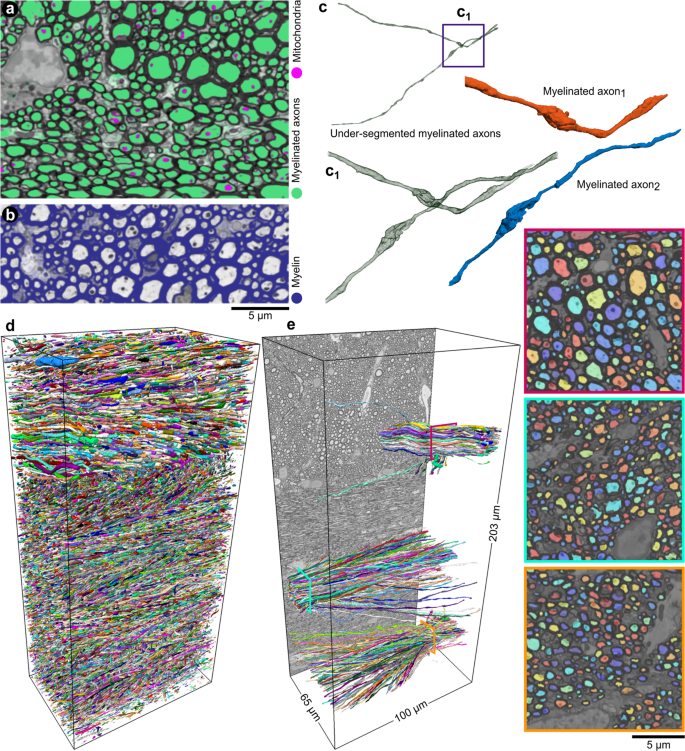
DeepACSON automated segmentation of white matter in 3D electron microscopy

Magnetically-actuated microposts stimulate axon growth - ScienceDirect

Scanning electron microscopy (SEM) of the microscopic structure of the

Extremely Low Forces Induce Extreme Axon Growth
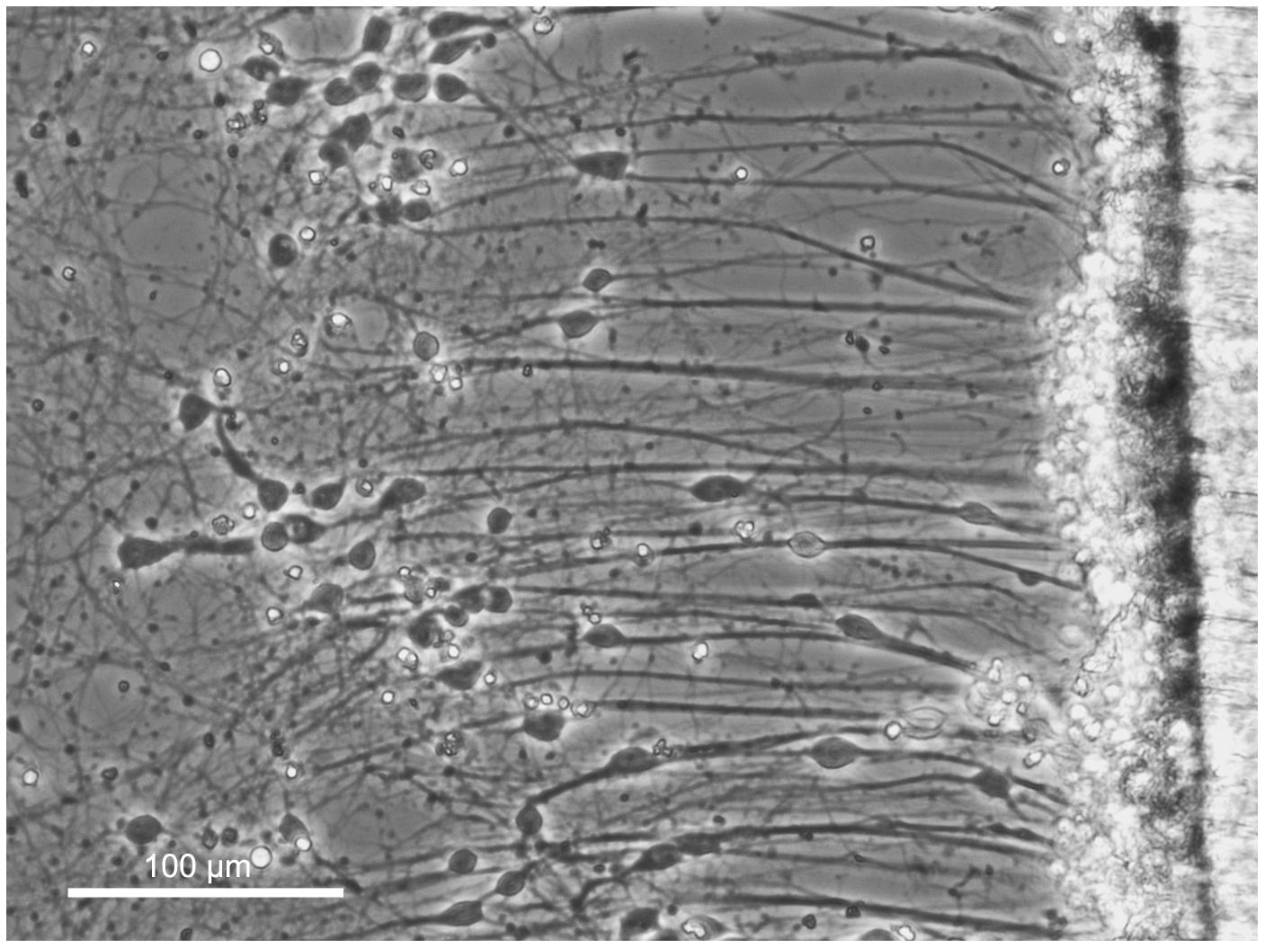
Cultured Axonal Injury, Smith Neurotrauma Lab
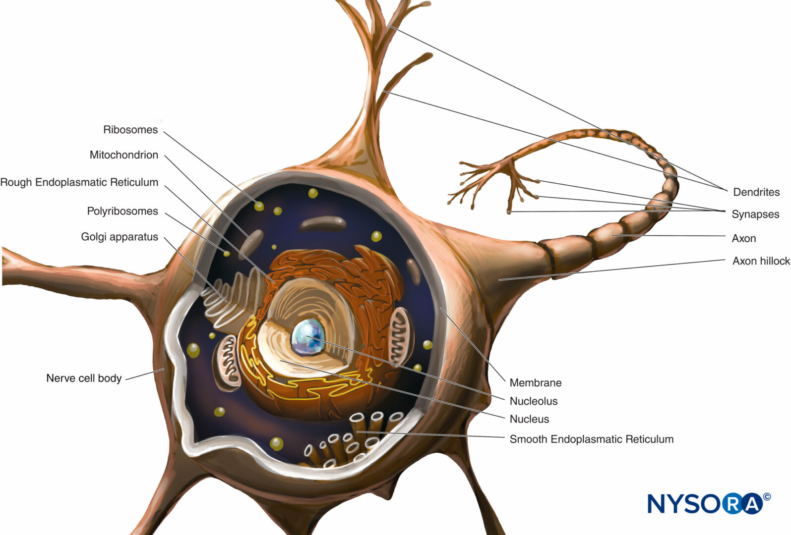
Histology of the Peripheral Nerves and Light Microscopy - NYSORA
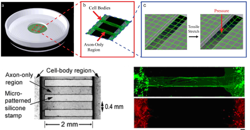
Frontiers Biomechanical Forces Regulate Gene Transcription During Stretch-Mediated Growth of Mammalian Neurons