Probe position and normal images obtained during E-FAST examination.
4.7 (186) · € 35.50 · En Stock

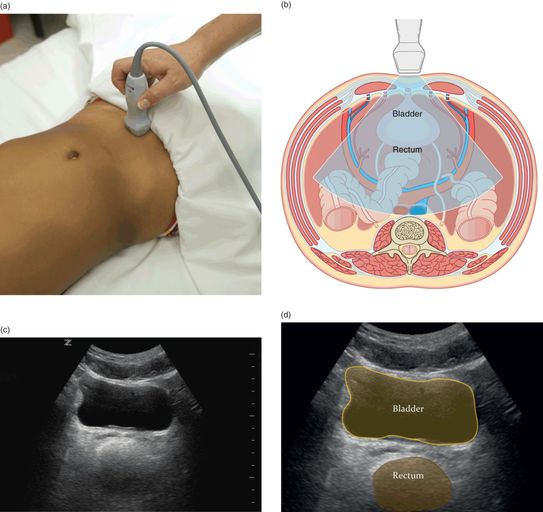
Download scientific diagram | Probe position and normal images obtained during E-FAST examination. (A) Right upper quadrant view demonstrating interface between liver and kidney (Morison's pouch). (B) Left upper quadrant view demonstrating spleen and kidney interface. (C) Left transverse view of the bladder. (D) Subcostal or subxiphoid view using the liver as a window to view the heart. (E) Anterior lung view. (F) Anterior lung view with US set to motion mode: this depicts a 1-dimensional view (marked on the top of the screen) as it changes over time (marked on the bottom of the screen); straight lines represent static soft tissue above the granular pattern representing the sliding of the pleura back and forth over time. E-FAST, Extended Focused Assessment with Sonography in Trauma examination. Used by permission from Introduction to Bedside Ultrasound, Vol 1, Dawson M, Mallin M, eds. Lexington, KY: Emergency Ultrasound Solutions; 2012: chap 1. from publication: Point-of-Care Ultrasound in Established Settings | The original and most widely accepted applications for point-of-care ultrasound (POCUS) are in the settings of trauma, shock, and bedside procedures. Trauma was the original setting for the introduction of POCUS and has been standardized under the four-plus view examination | Point-of-Care Systems, Ultrasound and Ultrasonography | ResearchGate, the professional network for scientists.

Focused Assessment with Sonography in Trauma (FAST) in 2017: What Radiologists Can Learn
Focused Assessment with Sonography in Trauma (FAST), Emergency and Clinical Ultrasound Board Review
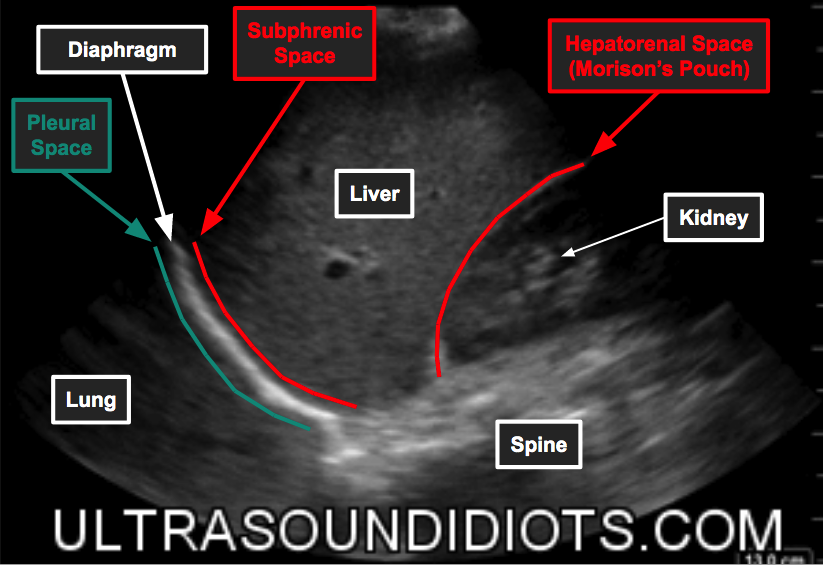
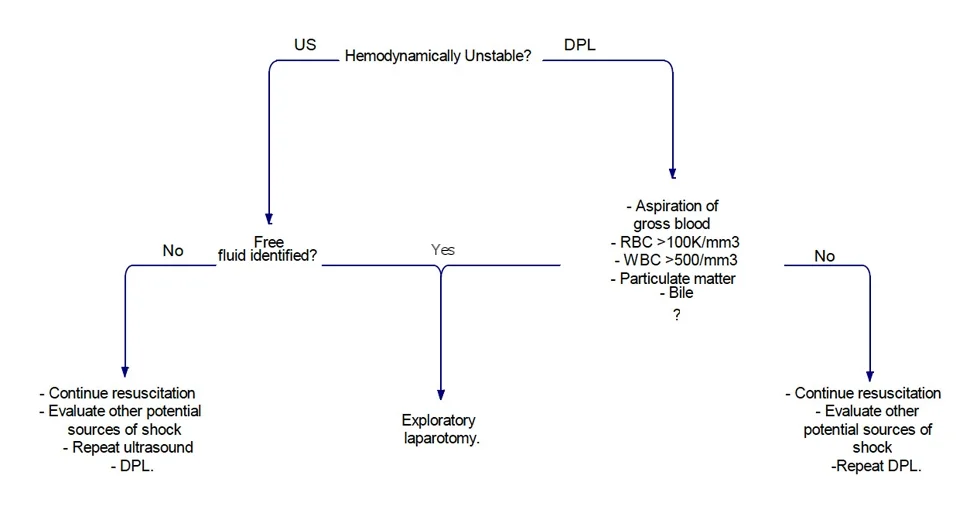
Ultrasound Idiots — Trauma / EFAST
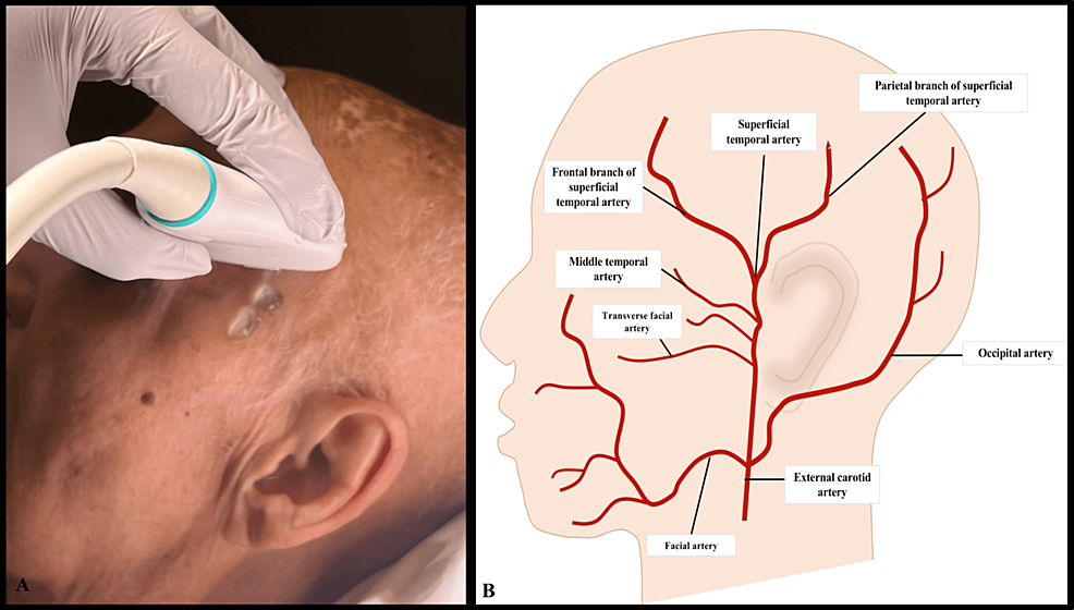
Cureus, Temporal Artery Ultrasound for the Diagnosis of Giant Cell Arteritis in the Emergency Department
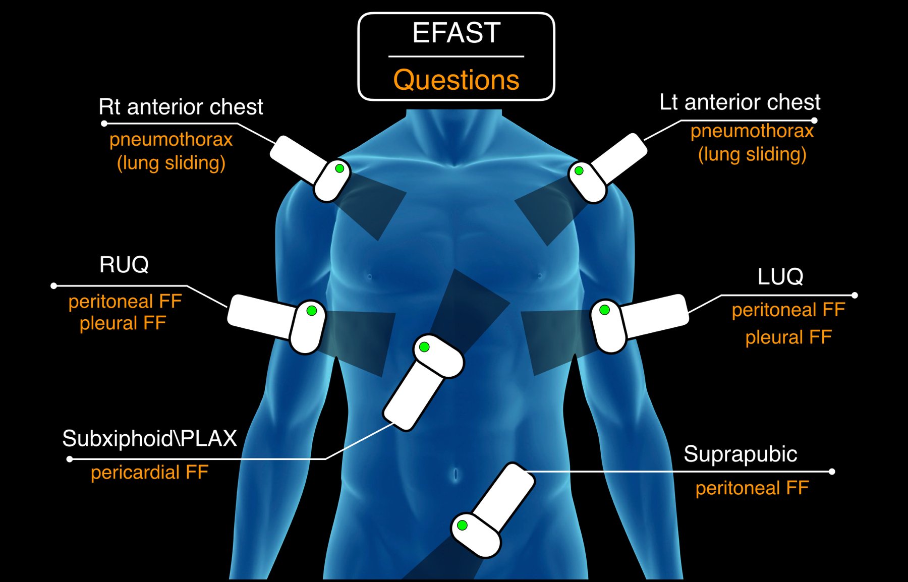
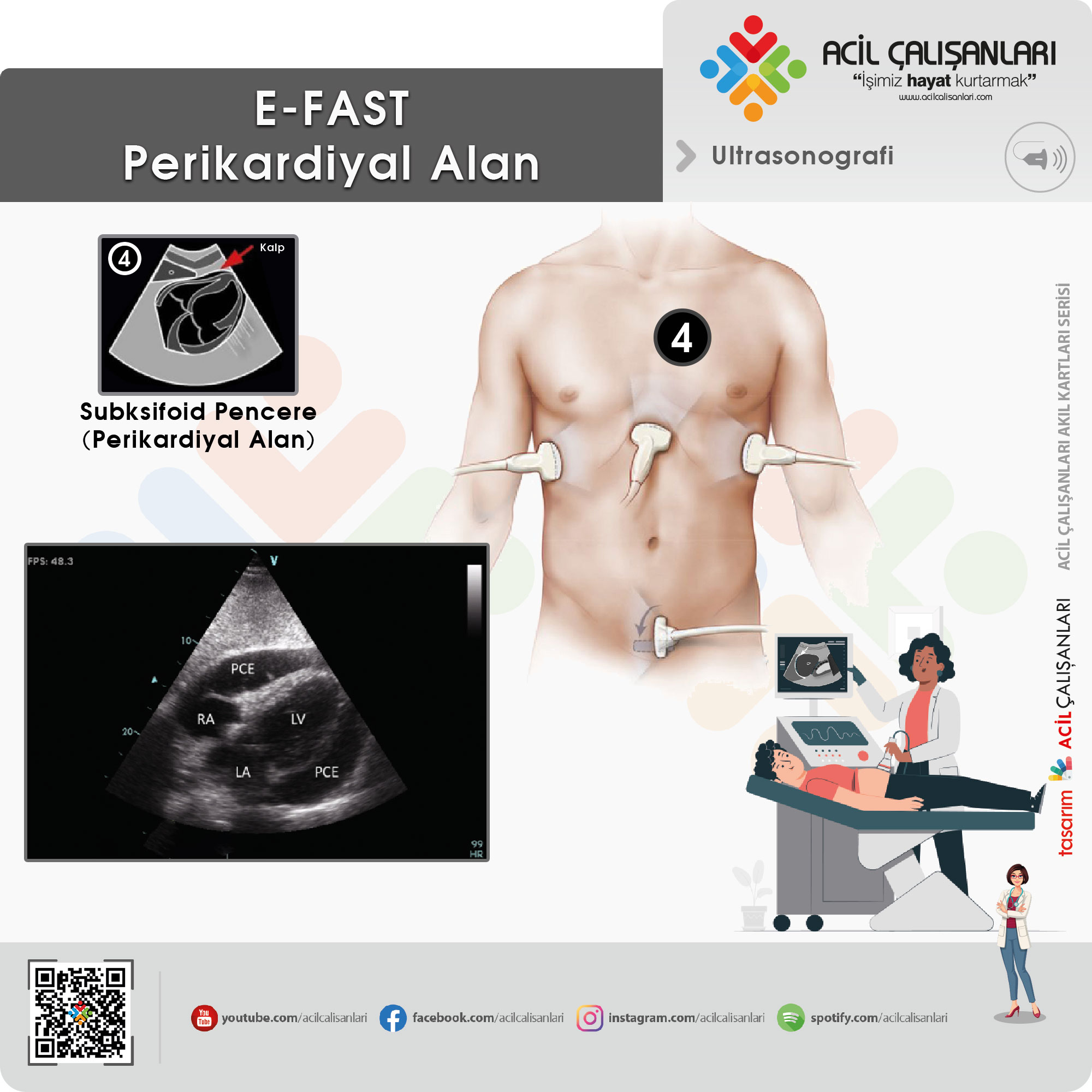
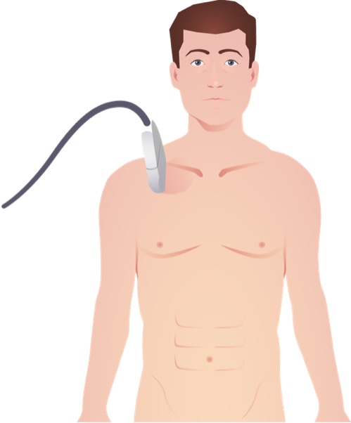
Probe placement for e-FAST: (1) subxiphoid (pericardial window); (2)
Scanning School - FAST and eFAST — Taming the SRU
Focused Assessment with Sonography for Trauma (FAST)

A, Probe position for Morison's pouch (RUQ). B, Sonographic view of the
A Beginner's Guide to Ultrasound, FAST scan

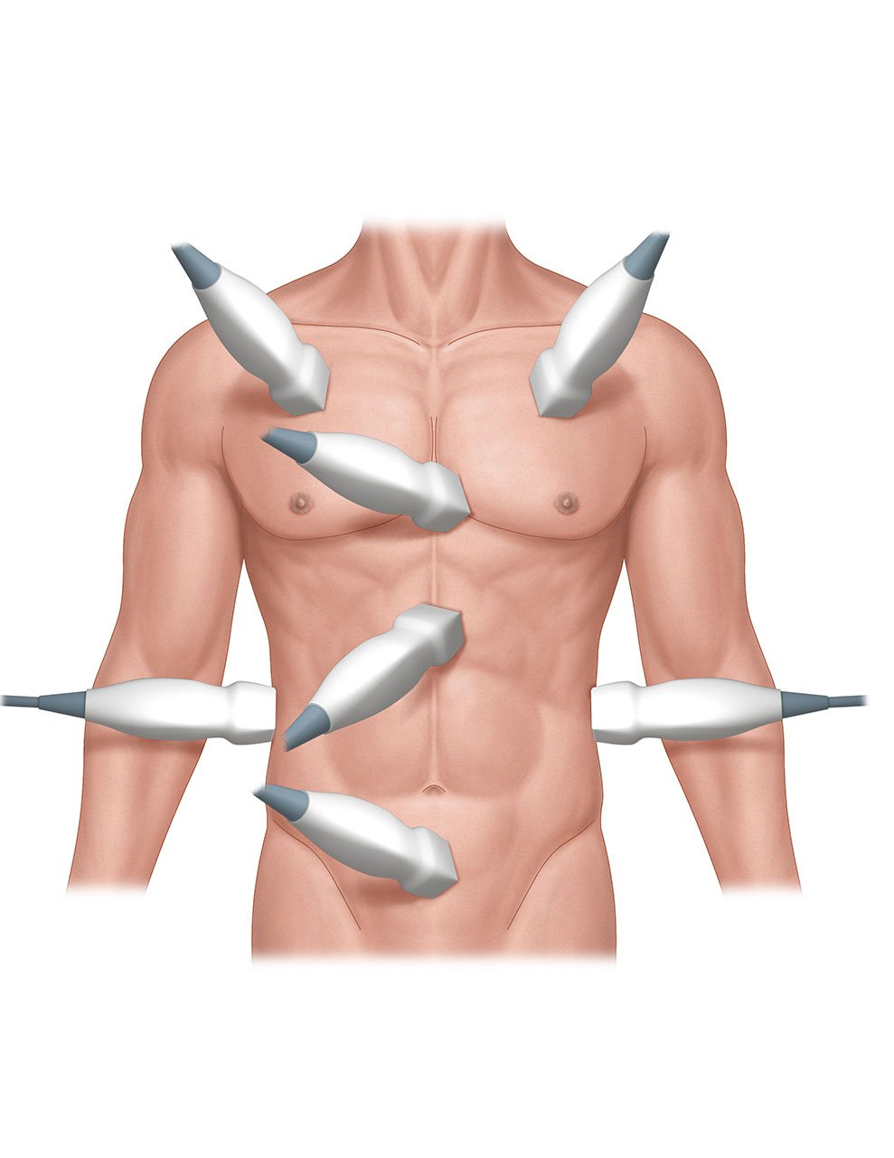
A: Essential probe positions for the EFAST exam Pericardium: The most
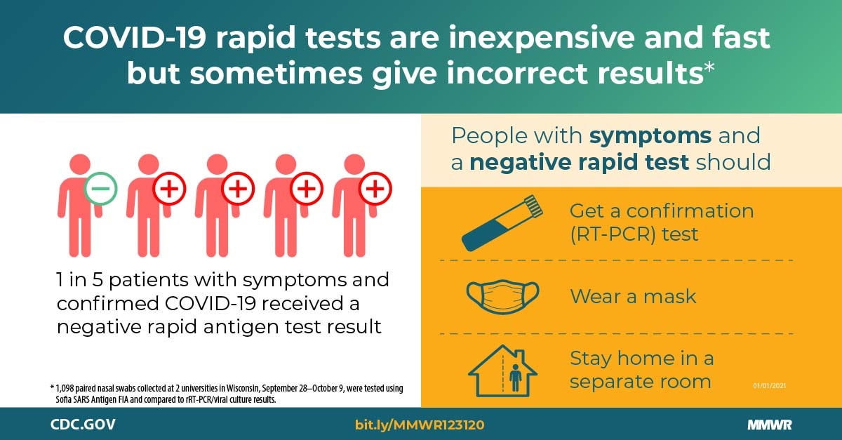
Performance of an Antigen-Based Test for Asymptomatic and Symptomatic SARS-CoV-2 Testing at Two University Campuses — Wisconsin, September–October 2020
E-Fast

How To Do E-FAST Examination - Critical Care Medicine - Merck Manuals Professional Edition
The extended focused assessment with sonography for trauma (E-FAST)

FAST Sonoguide
/product/86/0215/1.jpg?0263)